
Managing head & neck care pathway with ultrasound
Improve your thyroid workflow & increase clinical confidence with
Managing head & neck care pathway with ultrasound
Improve your thyroid workflow & increase clinical confidence with AI powered solutions
Detection
Clinical challenges
While ultrasound is a powerful tool for detecting thyroid diseases, clinicians do face several challenges: Classification of nodules, multiple nodule examinations & reproducibility of the images. Access to advanced ultrasound techniques & features can provide additional information about thyroid nodules. Ultrasound can leverage AI to aid in decision support, workflow efficiency and reproducibility of examination.
The incidence of thyroid nodules in the population rises year by year, and ultrasound is an important methodology that is currently used in the evaluation of the thyroid gland because it is noninvasive, safe, and economical. However, ultrasonic images may have disadvantages, such as relatively lower contrast resolution compare to other modality, time consuming and heavily dependent on operator interpretation. Access too advanced ultrasound techniques and features can provide additional information about thyroid nodule and potentially overcome these limitation.
The incidence of thyroid nodules in the population rises year by year, and ultrasound is an important methodology that is currently used in the evaluation of the thyroid gland because it is noninvasive, safe, and economical. However, ultrasonic images may have disadvantages, such as relatively lower contrast resolution compare to other modality, time consuming and heavily dependent on operator interpretation. Access too advanced ultrasound techniques and features can provide additional information about thyroid nodule and potentially overcome these limitation.
Solutions
Probes powered by XDclear™ technology, high quality B mode imaging, sensitive flow modes & AI powered solutions support clinicians to feel confident within the complexity of thyroid imaging & disease detection.

A probe solution for every need
Dedicated probes can help improve the confidence. XDclear™ technology helps increase penetration and simultaneously delivers high-definition resolution throughout the image.
LOGIQ Ultrasound have a portfolio of transducers with latest innovations that includes high frequency probes, from large footprints to light hockey sticks, high performing low frequency, extended bandwidths and sensor embedded solutions for navigation.
LOGIQ Ultrasound have a portfolio of transducers with latest innovations that includes high frequency probes, from large footprints to light hockey sticks, high performing low frequency, extended bandwidths and sensor embedded solutions for navigation.
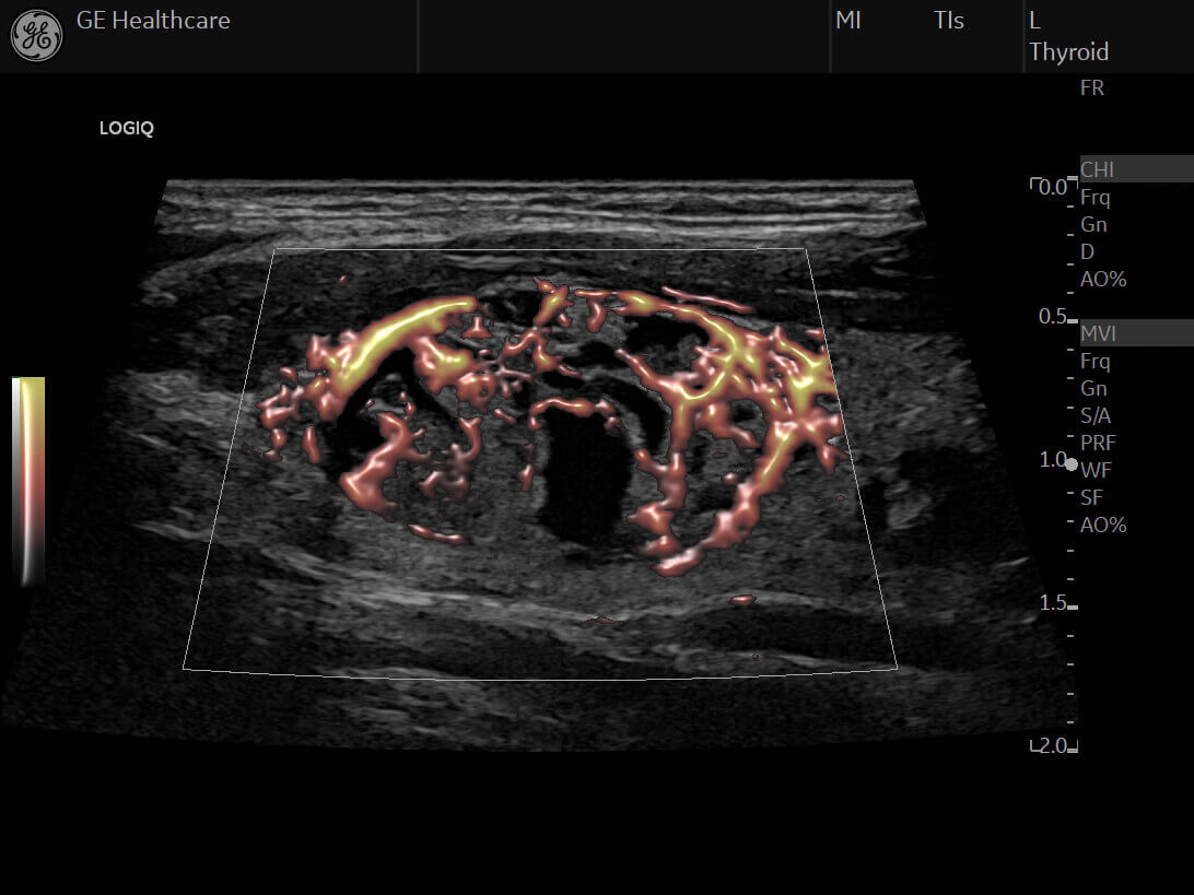
B mode Image Quality with cSound™ Image former delivers a focus by pixel combined with the new Advanced SRI to elevate image quality improving diagnostic confidence. High-definition flow modes, as Micro Vascular Imaging (MVI) with Radiantflow™ are able to help detect and display micro vessels and show their clear interconnection helping increase clinical confidence:
- a clearer definition of the vessels with Radiantflow™,
- an easier display of interconnecting smaller vessels; detecting more vascularity than Color Doppler and Power Doppler technologies,
- a better detection of subtle abnormality in traditionally “blind zones”, including thyroid capsular tumor involvement or lymph node cortex neoangiogenesis
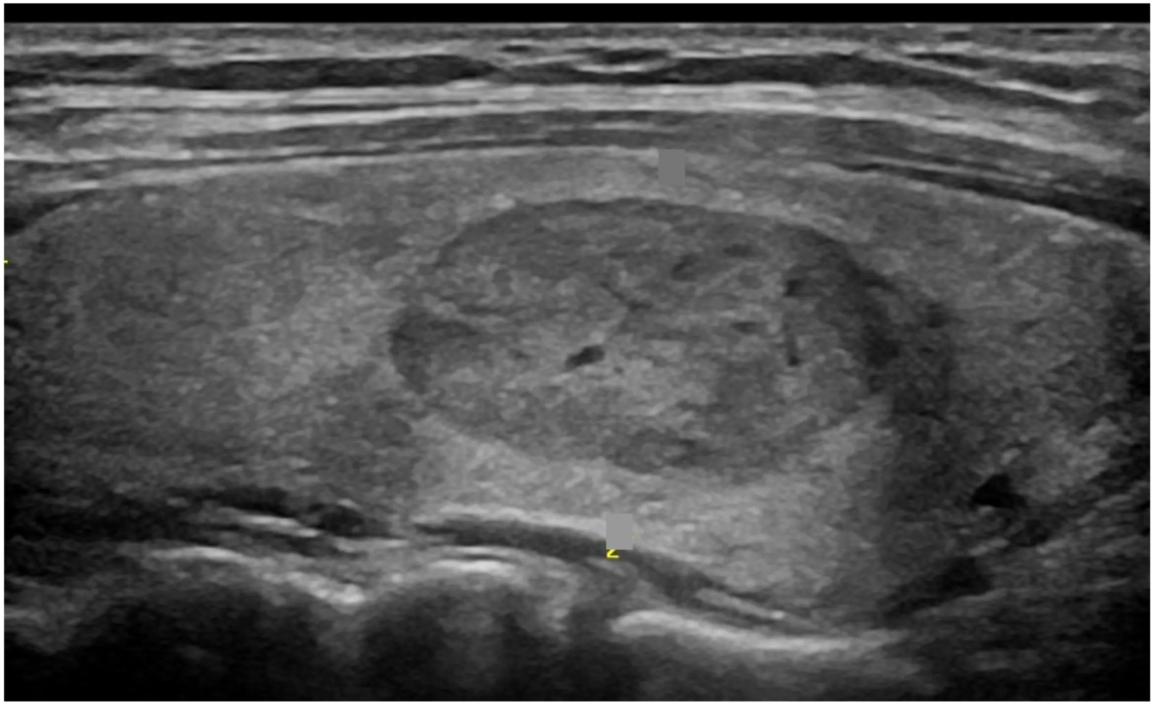
User identifies a thyroid nodule
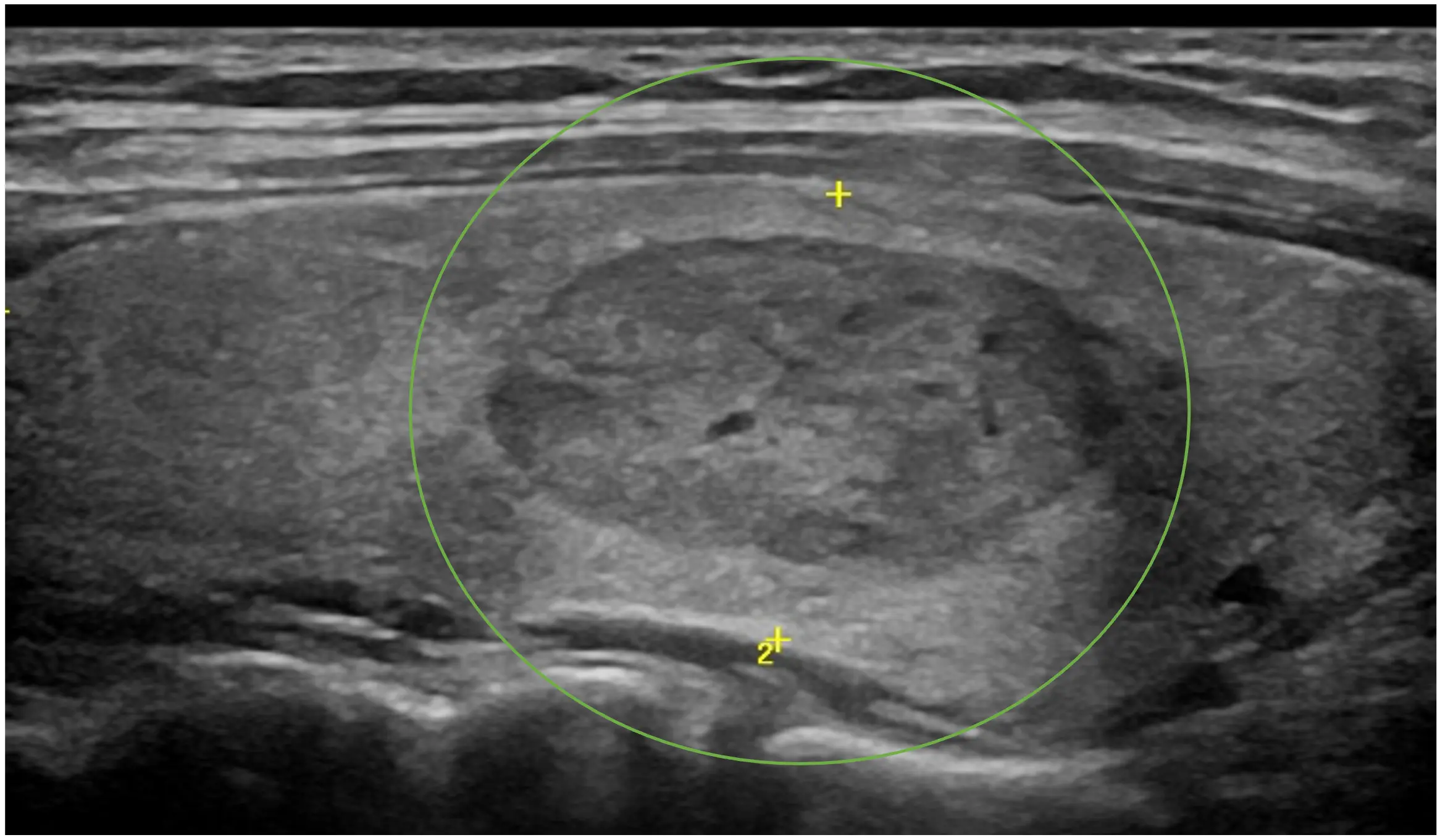
User clicks on the nodule and simply expands a graphical circle to encompass it

The algorithm segments the nodule, providing a trace and extents of the nodule
Harnessing the power of AI
This AI-based productivity tool automatically traces nodule boundaries and generates two-dimensional measurements with just a few keystrokes. User identifies a nodule with a single click and calipers are automatically placed to measure the nodule, saving keystrokes and providing consistency.
PIUR tUS inside is an AI based solution available in LOGIQ™ ultrasound devices, which adds a 3D layer to the 2D ultrasound images which previously could have only been generated using other 3D imaging technologies like CT or MRI. This solution offers improvements in today’s challenging and timely thyroid workflow by reducing scanning & reporting time.
A magnetically attached sensor on supported transducers ML6-15-D, ML4-20-D, L3-12-D detects the users scan movement and allows to shorten the conventional imaging workflow to a single sweep per lobe side.
Learn more about the solution A magnetically attached sensor on supported transducers ML6-15-D, ML4-20-D, L3-12-D detects the users scan movement and allows to shorten the conventional imaging workflow to a single sweep per lobe side.
High-performace Probes
A probe solution for every need
Dedicated probes can help improve the confidence. XDclear™ technology helps increase penetration and simultaneously delivers high-definition resolution throughout the image.
LOGIQ Ultrasound have a portfolio of transducers with latest innovations that includes high frequency probes, from large footprints to light hockey sticks, high performing low frequency, extended bandwidths and sensor embedded solutions for navigation.
LOGIQ Ultrasound have a portfolio of transducers with latest innovations that includes high frequency probes, from large footprints to light hockey sticks, high performing low frequency, extended bandwidths and sensor embedded solutions for navigation.
B Mode & Flow Modes
B mode Image Quality with cSound™ Image former delivers a focus by pixel combined with the new Advanced SRI to elevate image quality improving diagnostic confidence. High-definition flow modes, as Micro Vascular Imaging (MVI) with Radiantflow™ are able to help detect and display micro vessels and show their clear interconnection helping increase clinical confidence:
- a clearer definition of the vessels with Radiantflow™,
- an easier display of interconnecting smaller vessels; detecting more vascularity than Color Doppler and Power Doppler technologies,
- a better detection of subtle abnormality in traditionally “blind zones”, including thyroid capsular tumor involvement or lymph node cortex neoangiogenesis
Auto Lesion Segmentation
User identifies a thyroid nodule
User clicks on the nodule and simply expands a graphical circle to encompass it
The algorithm segments the nodule, providing a trace and extents of the nodule
Harnessing the power of AI
This AI-based productivity tool automatically traces nodule boundaries and generates two-dimensional measurements with just a few keystrokes. User identifies a nodule with a single click and calipers are automatically placed to measure the nodule, saving keystrokes and providing consistency.
PIUR tUS inside
PIUR tUS inside is an AI based solution available in LOGIQ™ ultrasound devices, which adds a 3D layer to the 2D ultrasound images which previously could have only been generated using other 3D imaging technologies like CT or MRI. This solution offers improvements in today’s challenging and timely thyroid workflow by reducing scanning & reporting time.
A magnetically attached sensor on supported transducers ML6-15-D, ML4-20-D, L3-12-D detects the users scan movement and allows to shorten the conventional imaging workflow to a single sweep per lobe side.
Learn more about the solution A magnetically attached sensor on supported transducers ML6-15-D, ML4-20-D, L3-12-D detects the users scan movement and allows to shorten the conventional imaging workflow to a single sweep per lobe side.
Diagnosis
Clinical challenges
Timely and accurate diagnosis of thyroid nodule is essential for appropriate treatment. However measuring and interpreting thyroid nodules are time and resource-consuming processes in ultrasound. Despite the ACR®Thyroid Imaging Reporting and Data System (TI-RADS™),characterization is often inconsistent from clinician to clinician and may lead to unnecessary biopsies and overtreatment with insignificant clinical outcome.
Solutions
In addition to B-Mode and various Flow Modes, a comprehensive solutions is available to increases decision-making confidence in the diagnosis of thyroid nodule, including Thyroid Assistant, powered by Koios DS™, 2D shearwave elastography, contrast enhanced ultrasound and B-steer+.

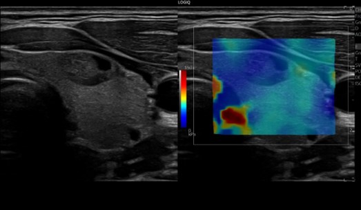
Micro-vascular Imaging (MVI) & Flow Modes
High-definition flow modes, as Micro Vascular Imaging (MVI) with Radiantflow™ are able to help detect and display identify tiny vessels in lesions and lymph nodes and show their clear interconnection helping increase clinical confidence:
- a clearer definition of the vessels with Radiantflow™,
- an easier display of interconnecting smaller vessels; detecting more vascularity than Color Doppler and Power Doppler technologies,
- a better detection of subtle abnormality in traditionally “blind zones”, including thyroid capsular tumor involvement or lymph node cortex neoangiogenesis
2D Shear Wave Elastography
Elevate Diagnostic Confidence
Enables non-invasive assessment of lesion stiffness in thyroid with a Shear Wave Elastography Quality Indicator to increase the confidence in the characterization of lesions (as benign or malignant). Strain elastography is also available for semiquantitative tissue assessment.
Enables non-invasive assessment of lesion stiffness in thyroid with a Shear Wave Elastography Quality Indicator to increase the confidence in the characterization of lesions (as benign or malignant). Strain elastography is also available for semiquantitative tissue assessment.

B-STEER+
Enhanced needle visualization for higher confidence
B-Steer+ enables enhanced visualization of the needles structure during interventional procedures, helping improve user confidence. (biopsy procedure)
Highlights
B-Steer+ enables enhanced visualization of the needles structure during interventional procedures, helping improve user confidence. (biopsy procedure)
Highlights
- Up to 12 selectable steering angles available (six each direction)
- Separate gain control for needle refleciton
- Available on all linear transducers
- Quick one-button operation

CEUS
Elevate Diagnostic confidence
Contrast-Enhanced Ultrasound helps to get a clear picture of tissue structure and suspicious mass vascularity by optimizing the balance between penetration and resolution for improved contrast sensitivity.
Contrast-Enhanced Ultrasound helps to get a clear picture of tissue structure and suspicious mass vascularity by optimizing the balance between penetration and resolution for improved contrast sensitivity.

Thyroid Assistant, powered by Koios DS™
Thyroid Assistant, powered by Koios DS™, is a clinical decision support software that automatically populates all TI-RADS descriptors and offers an AI-based cancer risk assessment. It also makes a recommendation on whether the patient has benign nodules or is a candidate for Fine Needle Aspiration (FNA) or follow-up monitoring. This tool supports to reduce variability in TI-RADS categorization and increase decision-making confidence and speed.
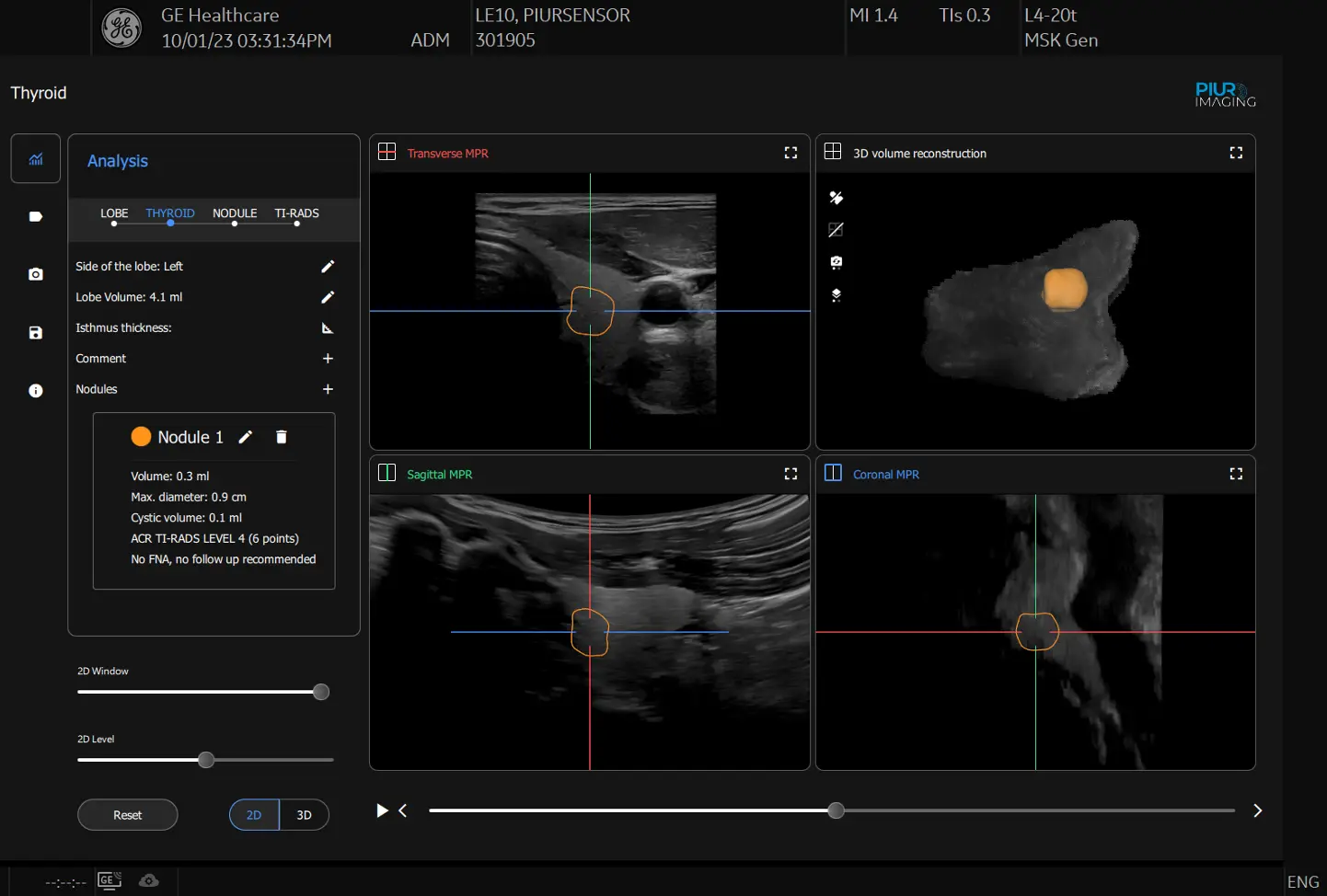
PIUR tUS inside
PIUR tUS inside is an AI based solution available in LOGIQ™ ultrasound devices, which adds a 3D layer to the 2D ultrasound images which previously could have only been generated using other 3D imaging technologies like CT or MRI. This solution offers improvements in today’s challenging and timely thyroid workflow by reducing scanning & reporting time.
A magnetically attached sensor on supported transducers ML6-15-D, ML4-20-D, L3-12-D detects the users scan movement and allows to shorten the conventional imaging workflow to a single sweep per lobe side.
Using AI algorithms, the thyroid analysis is performed on the reconstructed images, which automatically identify the lobe side and calculate the volume of the detected lobe. Based on one-click detected nodules, ACR TI-RADS™ classification is provided through semi-automated AI, assisting the user in the clinical recommendation for the analyzed nodule.
MVI & Flow Modes
Micro-vascular Imaging (MVI) & Flow Modes
High-definition flow modes, as Micro Vascular Imaging (MVI) with Radiantflow™ are able to help detect and display identify tiny vessels in lesions and lymph nodes and show their clear interconnection helping increase clinical confidence:
- a clearer definition of the vessels with Radiantflow™,
- an easier display of interconnecting smaller vessels; detecting more vascularity than Color Doppler and Power Doppler technologies,
- a better detection of subtle abnormality in traditionally “blind zones”, including thyroid capsular tumor involvement or lymph node cortex neoangiogenesis
2D Shear Wave Elastography
2D Shear Wave Elastography
Elevate Diagnostic Confidence
Enables non-invasive assessment of lesion stiffness in thyroid with a Shear Wave Elastography Quality Indicator to increase the confidence in the characterization of lesions (as benign or malignant). Strain elastography is also available for semiquantitative tissue assessment.
Enables non-invasive assessment of lesion stiffness in thyroid with a Shear Wave Elastography Quality Indicator to increase the confidence in the characterization of lesions (as benign or malignant). Strain elastography is also available for semiquantitative tissue assessment.
B-STEER+
B-STEER+
Enhanced needle visualization for higher confidence
B-Steer+ enables enhanced visualization of the needles structure during interventional procedures, helping improve user confidence. (biopsy procedure)
Highlights
B-Steer+ enables enhanced visualization of the needles structure during interventional procedures, helping improve user confidence. (biopsy procedure)
Highlights
- Up to 12 selectable steering angles available (six each direction)
- Separate gain control for needle refleciton
- Available on all linear transducers
- Quick one-button operation
CEUS
CEUS
Elevate Diagnostic confidence
Contrast-Enhanced Ultrasound helps to get a clear picture of tissue structure and suspicious mass vascularity by optimizing the balance between penetration and resolution for improved contrast sensitivity.
Contrast-Enhanced Ultrasound helps to get a clear picture of tissue structure and suspicious mass vascularity by optimizing the balance between penetration and resolution for improved contrast sensitivity.
Thyroid Assistant, powered by Koios DS™
Thyroid Assistant, powered by Koios DS™
Thyroid Assistant, powered by Koios DS™, is a clinical decision support software that automatically populates all TI-RADS descriptors and offers an AI-based cancer risk assessment. It also makes a recommendation on whether the patient has benign nodules or is a candidate for Fine Needle Aspiration (FNA) or follow-up monitoring. This tool supports to reduce variability in TI-RADS categorization and increase decision-making confidence and speed.
PIUR tUS inside
PIUR tUS inside
PIUR tUS inside is an AI based solution available in LOGIQ™ ultrasound devices, which adds a 3D layer to the 2D ultrasound images which previously could have only been generated using other 3D imaging technologies like CT or MRI. This solution offers improvements in today’s challenging and timely thyroid workflow by reducing scanning & reporting time.
A magnetically attached sensor on supported transducers ML6-15-D, ML4-20-D, L3-12-D detects the users scan movement and allows to shorten the conventional imaging workflow to a single sweep per lobe side.
Using AI algorithms, the thyroid analysis is performed on the reconstructed images, which automatically identify the lobe side and calculate the volume of the detected lobe. Based on one-click detected nodules, ACR TI-RADS™ classification is provided through semi-automated AI, assisting the user in the clinical recommendation for the analyzed nodule.
A magnetically attached sensor on supported transducers ML6-15-D, ML4-20-D, L3-12-D detects the users scan movement and allows to shorten the conventional imaging workflow to a single sweep per lobe side.
Using AI algorithms, the thyroid analysis is performed on the reconstructed images, which automatically identify the lobe side and calculate the volume of the detected lobe. Based on one-click detected nodules, ACR TI-RADS™ classification is provided through semi-automated AI, assisting the user in the clinical recommendation for the analyzed nodule.
Treatment & Follow up
Clinical challenges
After diagnosis , the management of thyroid nodule requires follow up with ultrasound. However there is Inconsistent interobserver variability in measurement changes in non interventional nodule and assessing treatment efficacy post treatment. In cases with multiple nodule, this increases the complexity and accuracy of the evaluation.
Solutions
Reproducibility of imaging is the key to an effective follow up. Feature such as PIUR tUS inside and compare assist provide users with increased clinical confident during examination.
Ultrasound imaging in thyroid nodule diagnosis, therapy, and follow‐up: Current status and future trends - Boers - 2023:
Journal of Clinical Ultrasound - Wiley Online Library 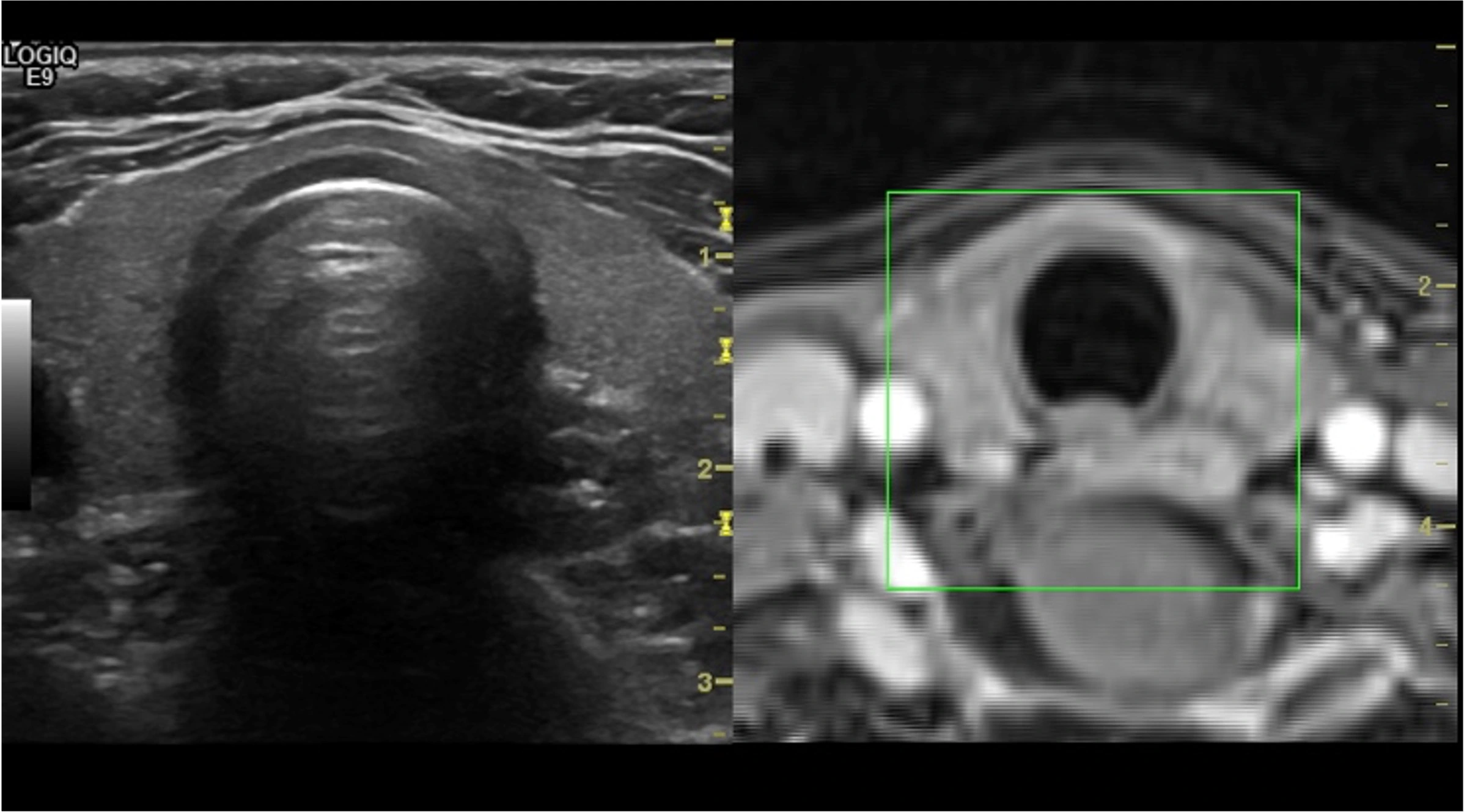
US/CT images of thyroid in treatment follow-up
Volume Navigation with Fusion Imaging & Needle Tracking
Merge real-time ultrasound with a volume DICOM® MR/CT dataset to facilitate fusion guidance of biopsies/RFA of masses.
- Provides the advantages of US Real Time Imaging associated with High spatial/contrast of MRI, CT or Handheld SPECT Imaging
- Helps increase precision and accuracy of imaging-guided interventional procedures like biopsy
- Helps identify lesions poorly visualized with US thanks to GPS
- Adequate visible lesions with US, but coexisting with difficult targets
- Active Tracker for Quick US/MR Auto-Registration and breath compensation
Compare Assistant
Elevate exam & workflow efficiency
Enables clinicians to easily view a prior study – ultrasound, CT or MR – and compare with current images in real time via a split screen on the monitor, helping to improve confidence and exam efficiency
Enables clinicians to easily view a prior study – ultrasound, CT or MR – and compare with current images in real time via a split screen on the monitor, helping to improve confidence and exam efficiency
PIUR tUS inside
PIUR tUS inside is an AI based solution available in LOGIQ™ ultrasound devices, which adds a 3D layer to the 2D ultrasound images which previously could have only been generated using other 3D imaging technologies like CT or MRI. This solution offers improvements in today’s challenging and timely thyroid workflow by reducing scanning & reporting time.
A magnetically attached sensor on supported transducers ML6-15-D, ML4-20-D, L3-12-D detects the users scan movement and allows to shorten the conventional imaging workflow to a single sweep per lobe side.
Using AI algorithms, the thyroid analysis is performed on the reconstructed images, which automatically identify the lobe side and calculate the volume of the detected lobe. Based on one-click detected nodules, ACR TI-RADS™ classification is provided through semi-automated AI, assisting the user in the clinical recommendation for the analyzed nodule.
A magnetically attached sensor on supported transducers ML6-15-D, ML4-20-D, L3-12-D detects the users scan movement and allows to shorten the conventional imaging workflow to a single sweep per lobe side.
Using AI algorithms, the thyroid analysis is performed on the reconstructed images, which automatically identify the lobe side and calculate the volume of the detected lobe. Based on one-click detected nodules, ACR TI-RADS™ classification is provided through semi-automated AI, assisting the user in the clinical recommendation for the analyzed nodule.
Volume Navigation with Fusion Imaging & Needle Tracking
US/CT images of thyroid in treatment follow-up
Volume Navigation with Fusion Imaging & Needle Tracking
Merge real-time ultrasound with a volume DICOM® MR/CT dataset to facilitate fusion guidance of biopsies/RFA of masses.
- Provides the advantages of US Real Time Imaging associated with High spatial/contrast of MRI, CT or Handheld SPECT Imaging
- Helps increase precision and accuracy of imaging-guided interventional procedures like biopsy
- Helps identify lesions poorly visualized with US thanks to GPS
- Adequate visible lesions with US, but coexisting with difficult targets
- Active Tracker for Quick US/MR Auto-Registration and breath compensation
Compare Assistant
Compare Assistant
Elevate exam & workflow efficiency
Enables clinicians to easily view a prior study – ultrasound, CT or MR – and compare with current images in real time via a split screen on the monitor, helping to improve confidence and exam efficiency
Enables clinicians to easily view a prior study – ultrasound, CT or MR – and compare with current images in real time via a split screen on the monitor, helping to improve confidence and exam efficiency
PIUR tUS inside
PIUR tUS inside
PIUR tUS inside is an AI based solution available in LOGIQ™ ultrasound devices, which adds a 3D layer to the 2D ultrasound images which previously could have only been generated using other 3D imaging technologies like CT or MRI. This solution offers improvements in today’s challenging and timely thyroid workflow by reducing scanning & reporting time.
A magnetically attached sensor on supported transducers ML6-15-D, ML4-20-D, L3-12-D detects the users scan movement and allows to shorten the conventional imaging workflow to a single sweep per lobe side.
Using AI algorithms, the thyroid analysis is performed on the reconstructed images, which automatically identify the lobe side and calculate the volume of the detected lobe. Based on one-click detected nodules, ACR TI-RADS™ classification is provided through semi-automated AI, assisting the user in the clinical recommendation for the analyzed nodule.
A magnetically attached sensor on supported transducers ML6-15-D, ML4-20-D, L3-12-D detects the users scan movement and allows to shorten the conventional imaging workflow to a single sweep per lobe side.
Using AI algorithms, the thyroid analysis is performed on the reconstructed images, which automatically identify the lobe side and calculate the volume of the detected lobe. Based on one-click detected nodules, ACR TI-RADS™ classification is provided through semi-automated AI, assisting the user in the clinical recommendation for the analyzed nodule.
Back to Head & Neck care area site
Sind Sie an einem Angebot interessiert?
Jetzt Ihr LOGIQ konfigurieren

LOGIQ E10 Series

LOGIQ Fortis

LOGIQ P Series
Fragen?
Kontaktieren Sie uns
Kontakt 


