
Managing liver care with ultrasound
See and quantify liver disease
in a single exam
Managing liver care with ultrasound
See and quantify liver disease
in a single exam
Screening
Clinical challenges
While using ultrasound in the screening of high risk patients for liver Disease, there are some important signs to look at to perform both a morphological and a functional assessment. These signs include liver size, liver surface appearance or texture (e.g. for assessment of a potential fatty liver infiltration). Ultrasound can also spot potential liver lesions deserving a second level of diagnosis. Last, ultrasound is key to check the patency of the hepatic and portal veins.
Solutions
For a morphological and functional assessment of liver disease high quality B-Mode imaging, sensitive Flow Modes and non-invasive technique for steatosis grading are key in the screening stage.
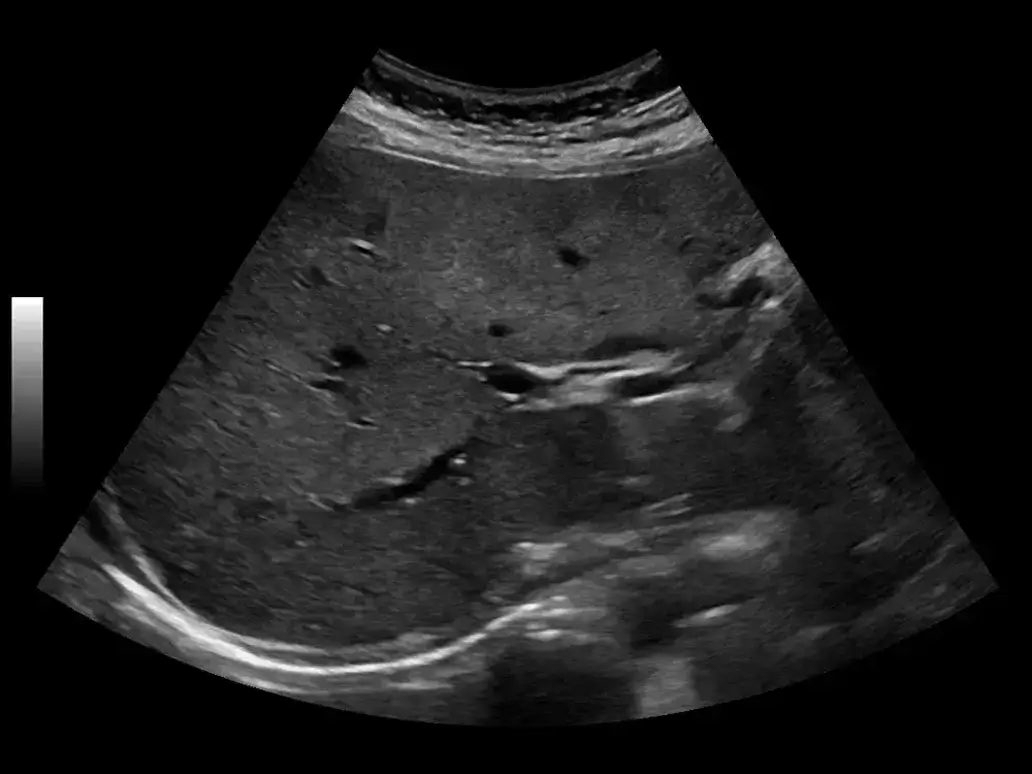
B-Mode for a morphological assessment
High quality resolution in B-Mode is the result of the latest Imaging processing techniques, as the cSound™ Imageformer and the most advanced probes technology. The result is an improved signal-to-noise ratio with outstanding image spatial, contrast resolution and uniformity. The images are often automatically optimized from near to far field.
Learn more about the advantages of the cSound™ imaging technology Learn more about the advantages of the XDclear™ Probe Technology 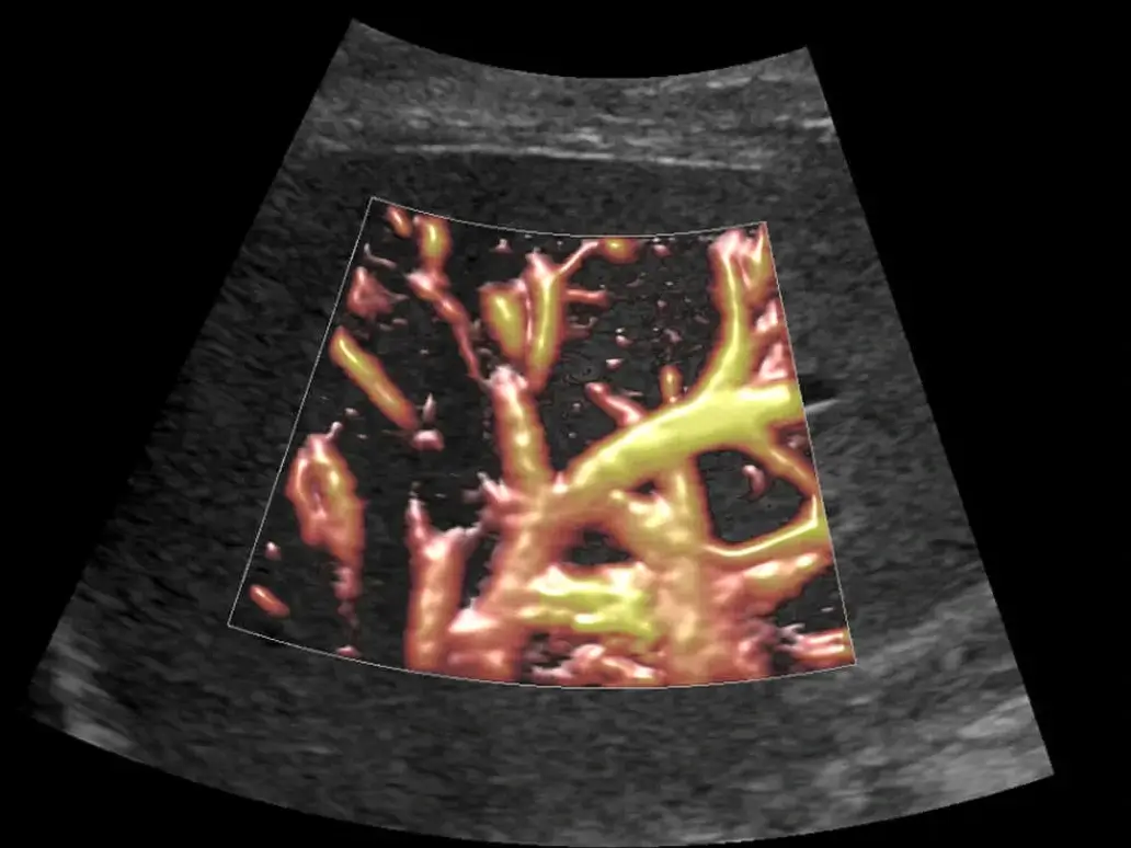
Flow Modes to check patency of Liver vasculature
The analysis of flows requires techniques with high resolution, sensitivity and accuracy in the delineation of vessels walls. Micro-vascular Imaging helps analyze slow flow states or small vessels in the Liver parenchyma or to study the structure of focal liver lesions. B-Flow, a Non-Doppler technique, can help see the real hemodynamics of the vessels in B-Mode, without the limitations of Doppler.
Learn more about the LOGIQ Vascular techniques 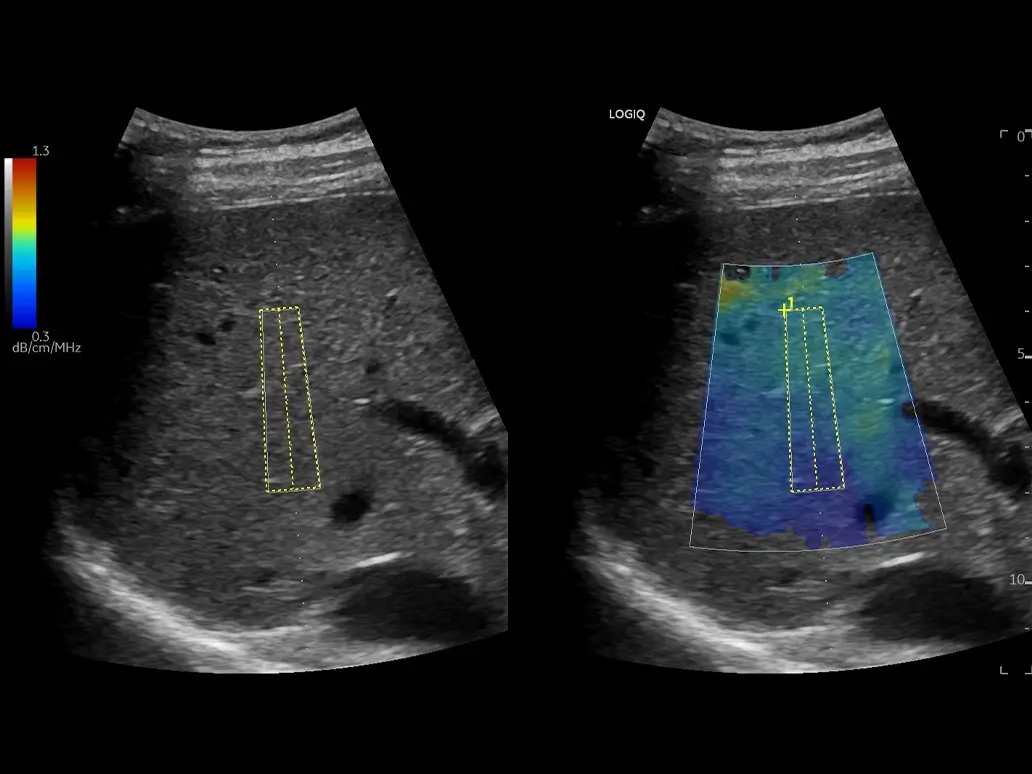
Ultrasound-Guided Attenuation Parameter (UGAP) for steatosis grading
An early assessment of fatty liver is possible either with a semi-qualitative assessment of B-Mode brightness comparing liver/kidney echo-levels or using a non-invasive quantitative tool as the Ultrasound-Guided Attenuation Parameter (UGAP). This easy technique results in a number associated with the degree of fatty liver, validated with the existing gold standards.
Learn how UGAP can help in steatosis grading B-Mode
B-Mode for a morphological assessment
High quality resolution in B-Mode is the result of the latest Imaging processing techniques, as the cSound™ Imageformer and the most advanced probes technology. The result is an improved signal-to-noise ratio with outstanding image spatial, contrast resolution and uniformity. The images are often automatically optimized from near to far field.
Learn more about the advantages of the cSound™ imaging technology Learn more about the advantages of the XDclear™ Probe Technology Flow Modes
Flow Modes to check patency of Liver vasculature
The analysis of flows requires techniques with high resolution, sensitivity and accuracy in the delineation of vessels walls. Micro-vascular Imaging helps analyze slow flow states or small vessels in the Liver parenchyma or to study the structure of focal liver lesions. B-Flow, a Non-Doppler technique, can help see the real hemodynamics of the vessels in B-Mode, without the limitations of Doppler.
Learn more about the LOGIQ Vascular techniques Steatosis grading with UGAP
Ultrasound-Guided Attenuation Parameter (UGAP) for steatosis grading
An early assessment of fatty liver is possible either with a semi-qualitative assessment of B-Mode brightness comparing liver/kidney echo-levels or using a non-invasive quantitative tool as the Ultrasound-Guided Attenuation Parameter (UGAP). This easy technique results in a number associated with the degree of fatty liver, validated with the existing gold standards.
Learn how UGAP can help in steatosis grading Diagnosis/Staging
Clinical challenges
In liver disease, diagnosis and staging is key to plan a personalized and effective therapy. Ultrasound is important in the management of patients with chronic liver disease, helping in steatosis grading and fibrosis/cirrhosis staging, in the study of the liver echo texture changes, as well as in the detection of ascites or in the diagnosis of portal hypertension with the measurements of spleen size and varices. In case of focal liver lesions, CEUS is the elected modality to detect small nodules and to perform their classification. In presence of HCC, primary cancer from a chronic liver condition, ultrasound can help in the diagnosis and treatment planning of the tumor.
Solutions
In addition to B-Mode and Flow Modes, a comprehensive package of solutions is available to support in the diagnosis and staging of liver disease including 2D Shear Wave Elastography, UGAP, Contrast-Enhanced Ultrasound and Volume Navigation.
B-Mode
Detect Liver echotexture changes: High quality resolution in B-Mode is the result of the latest imaging processing techniques, as the cSound Imageformer and the most advanced probes technology. The result is an improved signal-to-noise ratio with outstanding image spatial, contrast resolution and uniformity. The images are often automatically optimized from near to far field.
Learn more about the advantages of the cSound imaging technology Learn more about the advantages of the XDclear Probe Technology
Flow Modes to assess Liver vasculature
The comprehensive Flow Modes are important to study hepatic and portal veins, assess potential varices, as well as focal lesions vascular architecture and small capillaries.
Learn which is the LOGIQ Vascular solution fitting your needs 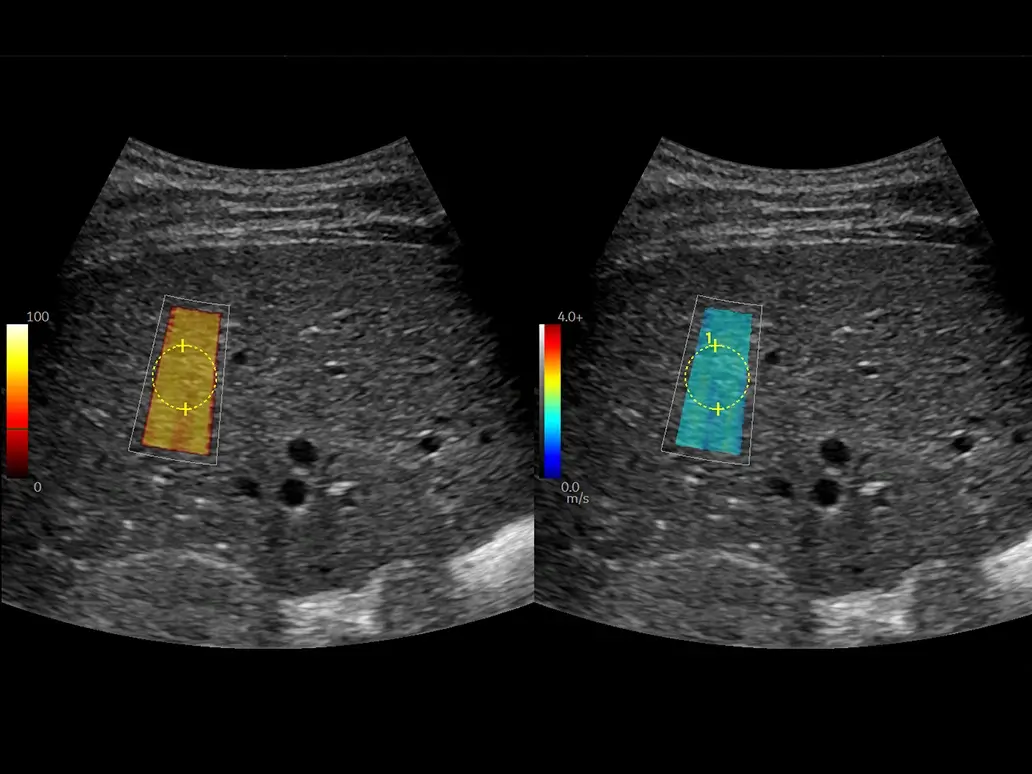
2D Shear Wave Elastography (2D SWE)
Liver fibrosis staging: 2D SWE is a non-invasive quantitative technique measuring the speed of propagation of Shear Waves in Liver, to assess the stiffness of the tissue. Quick, easy to use, intuitive, accurate and consistent, the LOGIQ 2D SWE is based on a proprietary technique. A quality Map is also available to increase user confidence.
Learn more about the LOGIQ 2D SWE technique Discover the latest 2D SWE LOGIQ Bibliography 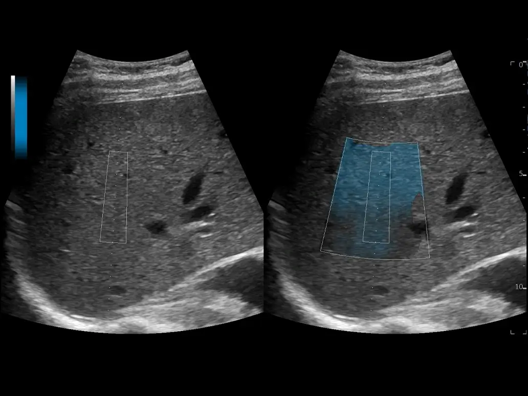
Ultrasound-Guided Attenuation Parameter (UGAP)
Non-invasive steatosis grading: An accurate assessment of fatty liver is possible using a non-invasive quantitative tool as the Ultrasound-Guided Attenuation Parameter (UGAP). This easy technique results in a number associated with the degree of fatty liver, validated with the existing gold standards.
Learn how you can benefit from LOGIQ UGAP technique in steatosis staging Discover the latest UGAP Bibliography 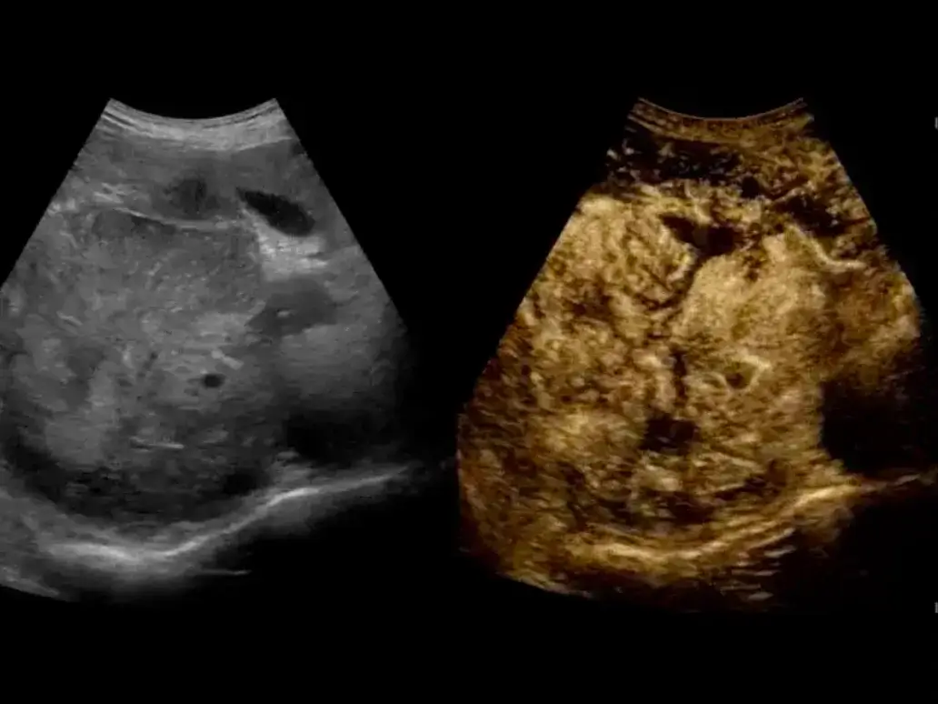
Contrast-Enhanced Ultrasound for FLLs
Detect and characterize Liver masses: CEUS are key in the detection and characterization of liver masses. With a high sensitivity, great penetration and low contrast need, LOGIQ Ultrasound provide a comprehensive package including integrated Parametric Imaging for contrast time arrival analysis and Time Intensity Curves.
Learn more about the LOGIQ CEUS capabilities 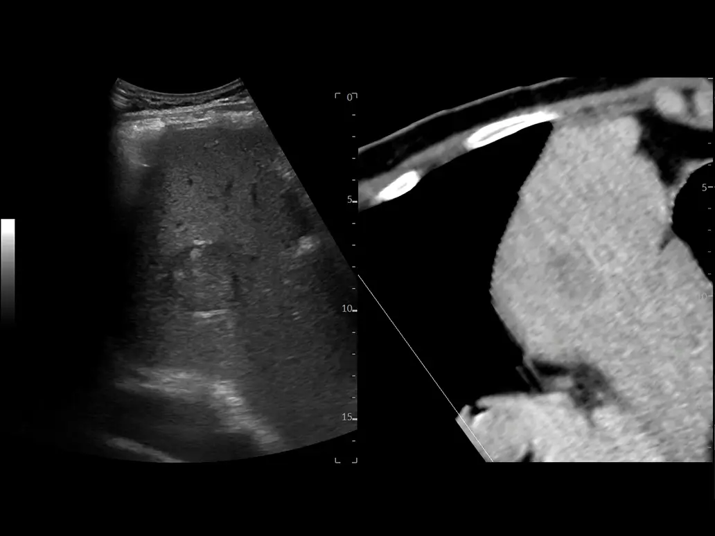
Volume Navigation with Fusion Imaging
Volume Navigation with Fusion Imaging provides a multi-modality approach to the study of Liver lesions combining the value of each modality as spatial, contrast and temporal resolution. It is key for the study of lesions hidden or partially visible in ultrasound. Easy to setup, with one-click registration technique and GE sensor-inside probes. It can work with 2D SWE, CEUS, and all vascular Modes.
B-Mode
B-Mode
Detect Liver echotexture changes: High quality resolution in B-Mode is the result of the latest imaging processing techniques, as the cSound Imageformer and the most advanced probes technology. The result is an improved signal-to-noise ratio with outstanding image spatial, contrast resolution and uniformity. The images are often automatically optimized from near to far field.
Learn more about the advantages of the cSound imaging technology Learn more about the advantages of the XDclear Probe Technology Flow Modes
Flow Modes to assess Liver vasculature
The comprehensive Flow Modes are important to study hepatic and portal veins, assess potential varices, as well as focal lesions vascular architecture and small capillaries.
Learn which is the LOGIQ Vascular solution fitting your needs 2D Shear Wave Elastography
2D Shear Wave Elastography (2D SWE)
Liver fibrosis staging: 2D SWE is a non-invasive quantitative technique measuring the speed of propagation of Shear Waves in Liver, to assess the stiffness of the tissue. Quick, easy to use, intuitive, accurate and consistent, the LOGIQ 2D SWE is based on a proprietary technique. A quality Map is also available to increase user confidence.
Learn more about the LOGIQ 2D SWE technique Discover the latest 2D SWE LOGIQ Bibliography Steatosis grading with UGAP
Ultrasound-Guided Attenuation Parameter (UGAP)
Non-invasive steatosis grading: An accurate assessment of fatty liver is possible using a non-invasive quantitative tool as the Ultrasound-Guided Attenuation Parameter (UGAP). This easy technique results in a number associated with the degree of fatty liver, validated with the existing gold standards.
Learn how you can benefit from LOGIQ UGAP technique in steatosis staging Discover the latest UGAP Bibliography Contrast-enhanced ultrasound (CEUS)
Contrast-Enhanced Ultrasound for FLLs
Detect and characterize Liver masses: CEUS are key in the detection and characterization of liver masses. With a high sensitivity, great penetration and low contrast need, LOGIQ Ultrasound provide a comprehensive package including integrated Parametric Imaging for contrast time arrival analysis and Time Intensity Curves.
Learn more about the LOGIQ CEUS capabilities Fusion Imaging
Volume Navigation with Fusion Imaging
Volume Navigation with Fusion Imaging provides a multi-modality approach to the study of Liver lesions combining the value of each modality as spatial, contrast and temporal resolution. It is key for the study of lesions hidden or partially visible in ultrasound. Easy to setup, with one-click registration technique and GE sensor-inside probes. It can work with 2D SWE, CEUS, and all vascular Modes.
Treatment
Clinical challenges
In focal liver disease, the role of Ultrasound is key in planning and guidance of treatments. In the TARGETING step, live ultrasound imaging is important to guide live biopsies, drive clips installation and plan the best trajectory to access the target in a interventional procedure. The challenges in interventional are the visibility of the lesion in ultrasound, the accessibility, the accuracy of the procedure and the safety of the patient. It is key to simplify the procedure and help in the management of emergency.
Solutions
In focal liver lesions treatment planning and guidance, accuracy and safety are key. Ultrasound can help being a real time imaging technique and having multiple tools to help increase confidence and act with speed when needed.
- Dedicated approach to liver biopsies and interventions with specialty probes
- Easy identification and targeting of FLLs with Contrast-Enhanced Ultrasound
- Increased clinical confidence with Fusion Imaging
Specialty probes for a dedicated approach
Dedicated probes can help improve the confidence in the targeting and interventional steps. LOGIQ Ultrasound have a portfolio of transducers with a small footprint for a better intercostal of subcostal approach as well as others dedicated to biopsy with a special shape allowing to avoid blind spots in the needle insertion. Easy to disinfect and be ready when needed. For a surgical approach, a T-shape probe with outstanding image quality and high CEUS performances is also available.
Contrast-Enhanced Ultrasound for FLLs
LOGIQ Ultrasound offer a comprehensive package of solutions to capture contrast agent's enhancement and process the information. In contrast imaging, the sensitivity of the system, the resolution of the image and the penetration are equally important. A combination of the three aspects is then what can make the difference. Time/Intensity curves or Parametric Imaging can be beneficial in this step of the care pathway.
Learn about the LOGIQ CEUS solutions
Volume Navigation with Fusion Imaging for increased confidence
Volume Navigation with Fusion Imaging provides a multi-modality approach to the study of Liver lesions combining the value of each modality as spatial, contrast and temporal resolution. Easy to setup, with one-click registration technique and unique GE sensor-inside probes, V Nav is quick, intuitive, consistent and accurate. It can work with 2D SWE, CEUS, and all vascular Modes. Available 2D & 3D GPS markers.
Learn more about the LOGIQ Interventional solutions Needle Tracking solutions for improved targeting
LOGIQ Ultrasound provides tools to draw the best needle trajectory prior to the intervention and track the needle path during the procedure. The Needle Tracking solutions allow to display the trajectory and the tip of the needle (either real or virtual) in real time, helping increase the confidence in the lesion targeting step of the procedure.
Learn more about the LOGIQ Interventional solutions Specialty Probes
Specialty probes for a dedicated approach
Dedicated probes can help improve the confidence in the targeting and interventional steps. LOGIQ Ultrasound have a portfolio of transducers with a small footprint for a better intercostal of subcostal approach as well as others dedicated to biopsy with a special shape allowing to avoid blind spots in the needle insertion. Easy to disinfect and be ready when needed. For a surgical approach, a T-shape probe with outstanding image quality and high CEUS performances is also available.
Contrast-Enhanced Ultrasound (CEUS)
Contrast-Enhanced Ultrasound for FLLs
LOGIQ Ultrasound offer a comprehensive package of solutions to capture contrast agent's enhancement and process the information. In contrast imaging, the sensitivity of the system, the resolution of the image and the penetration are equally important. A combination of the three aspects is then what can make the difference. Time/Intensity curves or Parametric Imaging can be beneficial in this step of the care pathway.
Learn about the LOGIQ CEUS solutions Fusion Imaging
Volume Navigation with Fusion Imaging for increased confidence
Volume Navigation with Fusion Imaging provides a multi-modality approach to the study of Liver lesions combining the value of each modality as spatial, contrast and temporal resolution. Easy to setup, with one-click registration technique and unique GE sensor-inside probes, V Nav is quick, intuitive, consistent and accurate. It can work with 2D SWE, CEUS, and all vascular Modes. Available 2D & 3D GPS markers.
Learn more about the LOGIQ Interventional solutions Needle Tracking
Needle Tracking solutions for improved targeting
LOGIQ Ultrasound provides tools to draw the best needle trajectory prior to the intervention and track the needle path during the procedure. The Needle Tracking solutions allow to display the trajectory and the tip of the needle (either real or virtual) in real time, helping increase the confidence in the lesion targeting step of the procedure.
Learn more about the LOGIQ Interventional solutions Follow up
Clinical challenges
During or after a treatment is important to monitor the efficacy of it, either in terms of response to a determined pharmacological treatment in case of CLD, check the success of a procedure (e.g. ablation) or follow up a long term anti-neo angiogenetic treatment in oncological patients. Monitoring is key to check if a change in the treatment is needed or a re-treatment is required.
Solutions
- Detect small changes in liver echotexture with B-Mode
- Assess the hepatic and portal veins status with Flow Modes
- Monitor any change in fibrosis staging with 2D Shear Wave Elastography
- Monitor any change in steatosis grading with UGAP
- Check the treatment results with Contrast-Enhanced Ultrasound
- Volume Navigation with Fusion Imaging
- Monitor the evolution of a disease with Compare Assistant
B-Mode for a morphological assessment
High quality resolution in B-Mode in monitoring is extremely important. In the LOGIQ ultrasound, it is the result of the latest imaging processing techniques, as the cSound™ Imageformer and the XDclear™ Architecture, with the most advanced XDclear™ probes technology. Binding multiple techniques, the result is an improved signal-to-noise ratio with outstanding image uniformity also when focuses do not require a manual adjustment and the image is always automatically optimized from near to far field. In treatment monitoring, you can well appreciate the changes of the liver pattern and the details of the areas that have been treated. High frequency Imaging is also adding much more details in case of superficial masses.
Learn more about the advantages of the cSound imaging technology Learn more about the advantages of the XDclear Probe Technology
Flow Modes for Liver vasculature
B-Flow is very helpful in the study of the Liver vessels. In some cases, it gives more information than standard Color techniques, being very sensitive displaying just the blood flows reflectors. More comprehensive Flow Modes are important to study portal vein, potential varices, as well as the vascular architecture changes during or after the treatments.
Learn more about the LOGIQ Vascular techniques
2D Shear Wave Elastography (2D SWE) for fibrosis staging
The monitoring of the response to treatment for patients with chronic liver disease can be done staging the Fibrosis and Cirrhosis using 2D Shear Wave Elastography, a non invasive quantitative technique measuring the speed of propagation of Shear Wave. Quick, easy to use, intuitive, accurate and consistent, the LOGIQ 2D SWE is based on a proprietary technique. A quality Map is also available to increase user confidence. In case of follow up of focal lesions ablation, it can help identify the real extension of the tissue necrosis and the presence of any viable tissue.
Learn more about the LOGIQ 2D SWE technique Discover the latest 2D SWE LOGIQ Bibliography
Ultrasound-Guided Attenuation Parameter (UGAP) for steatosis grading
LOGIQ Ultrasound provides tools to draw the best needle trajectory prior to the intervention and track the needle path during the procedure. The Needle Tracking solutions allow to display the trajectory and the tip of the needle (either real or virtual) in real time, helping increase the confidence in the lesion targeting step of the procedure.
Learn more about the LOGIQ UGAP technique Discover the latest LOGIQ UGAP Bibliography 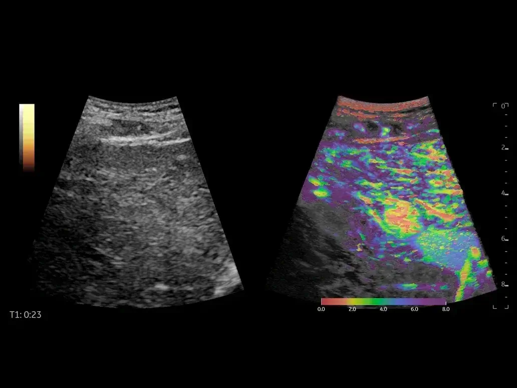
Contrast-Enhanced Ultrasound (CEUS) for monitoring
CEUS are key to check the success of focal lesions ablation, to monitor the final size of the ablated area and the potential presence of viable tissue. With a high sensitivity, great penetration and low contrast need, LOGIQ ultrasound provide a comprehensive package including integrated Parametric Imaging for contrast time arrival analysis and Time Intensity Curves.
Learn more about the LOGIQ CEUS solution
Volume Navigation with Fusion Imaging to check treatment success
Volume Navigation with Fusion Imaging provides a multi-modality approach to the study of Liver lesions combining the value of each modality as spatial, contrast and temporal resolution. After interventional procedures, as an ablation, V Nav allows to compare the US ablated area with CT/MRI dataset acquired prior and post the intervention and check the success. V Nav can work with 2D SWE, CEUS, and all vascular Modes that can help complete the analysis. Easy to setup, with one-click registration technique and GE sensor-inside probes, V Nav is quick, intuitive, consistent and accurate.
Learn more about the LOGIQ Interventional solutions B-Mode
B-Mode for a morphological assessment
High quality resolution in B-Mode in monitoring is extremely important. In the LOGIQ ultrasound, it is the result of the latest imaging processing techniques, as the cSound™ Imageformer and the XDclear™ Architecture, with the most advanced XDclear™ probes technology. Binding multiple techniques, the result is an improved signal-to-noise ratio with outstanding image uniformity also when focuses do not require a manual adjustment and the image is always automatically optimized from near to far field. In treatment monitoring, you can well appreciate the changes of the liver pattern and the details of the areas that have been treated. High frequency Imaging is also adding much more details in case of superficial masses.
Learn more about the advantages of the cSound imaging technology Learn more about the advantages of the XDclear Probe Technology Flow Modes
Flow Modes for Liver vasculature
B-Flow is very helpful in the study of the Liver vessels. In some cases, it gives more information than standard Color techniques, being very sensitive displaying just the blood flows reflectors. More comprehensive Flow Modes are important to study portal vein, potential varices, as well as the vascular architecture changes during or after the treatments.
Learn more about the LOGIQ Vascular techniques 2D Shear Wave Elastography
2D Shear Wave Elastography (2D SWE) for fibrosis staging
The monitoring of the response to treatment for patients with chronic liver disease can be done staging the Fibrosis and Cirrhosis using 2D Shear Wave Elastography, a non invasive quantitative technique measuring the speed of propagation of Shear Wave. Quick, easy to use, intuitive, accurate and consistent, the LOGIQ 2D SWE is based on a proprietary technique. A quality Map is also available to increase user confidence. In case of follow up of focal lesions ablation, it can help identify the real extension of the tissue necrosis and the presence of any viable tissue.
Learn more about the LOGIQ 2D SWE technique Discover the latest 2D SWE LOGIQ Bibliography UGAP
Ultrasound-Guided Attenuation Parameter (UGAP) for steatosis grading
LOGIQ Ultrasound provides tools to draw the best needle trajectory prior to the intervention and track the needle path during the procedure. The Needle Tracking solutions allow to display the trajectory and the tip of the needle (either real or virtual) in real time, helping increase the confidence in the lesion targeting step of the procedure.
Learn more about the LOGIQ UGAP technique Discover the latest LOGIQ UGAP Bibliography Contrast-Enhanced Ultrasound (CEUS)
Contrast-Enhanced Ultrasound (CEUS) for monitoring
CEUS are key to check the success of focal lesions ablation, to monitor the final size of the ablated area and the potential presence of viable tissue. With a high sensitivity, great penetration and low contrast need, LOGIQ ultrasound provide a comprehensive package including integrated Parametric Imaging for contrast time arrival analysis and Time Intensity Curves.
Learn more about the LOGIQ CEUS solution Fusion Imaging
Volume Navigation with Fusion Imaging to check treatment success
Volume Navigation with Fusion Imaging provides a multi-modality approach to the study of Liver lesions combining the value of each modality as spatial, contrast and temporal resolution. After interventional procedures, as an ablation, V Nav allows to compare the US ablated area with CT/MRI dataset acquired prior and post the intervention and check the success. V Nav can work with 2D SWE, CEUS, and all vascular Modes that can help complete the analysis. Easy to setup, with one-click registration technique and GE sensor-inside probes, V Nav is quick, intuitive, consistent and accurate.
Learn more about the LOGIQ Interventional solutions Back to liver care area site
Discover our clinical tutorials by care area
Go to clinical education

Interested in requesting a quotation?
Configure my LOGIQ

LOGIQ E10 Series

LOGIQ Fortis

LOGIQ P Series
Have a question?
We would love to hear from you.
Contact us 


